This idea has been awarded a VENI scholarship in the NWO talent scheme Veni, Vidi, Vici. Groot Jebbink was one of the 25 young scientists, less than 10 percent of all applicants within the ZonMW domain, receiving 250.000 euro for a three-year research program.
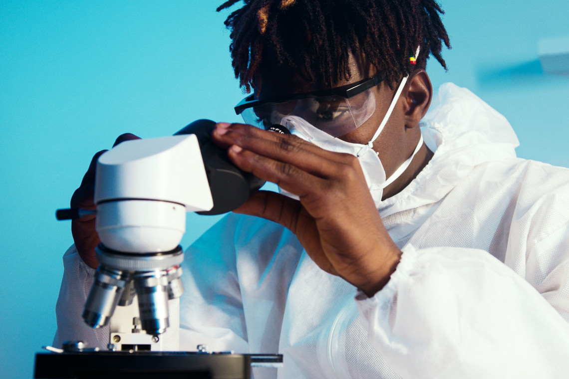
Erik Groot Jebbink can rightly be called a blood flow expert. For several years he has been using tiny gas bubbles, coated with a thin lipid layer for stabilization, to quantify local blood flow phenomena to improve the treatment of vascular diseases. Since a blood flow profile is closely related to potential vascular problems, this technique has resulted in an effective identification of problem areas and risk zones inside blood vessels, and potentially more effective long-term treatment of vascular diseases.
Cell-killing particles
But microbubbles can play a role in more medical fields. Groot Jebbink is now aiming for an improved treatment of another lethal disease: liver cancer. With the Veni scholarship, he wants to use his microbubble and blood flow experience to investigate if microbubbles can be used to improve the precision of delivering cell-killing radioactive Holmium particles to liver tumors.
Currently, liver tumors are treated palliatively with so-called radio-embolization techniques. Using this technique, a catheter is inserted into a large artery in the groin and moved all the way up to the liver. The tip of the catheter is then maneuvered as close as possible to the tumor, whereafter radioactive Holmium particles are injected. These have to find their way towards the tumor through smaller and smaller blood vessels, until they get stuck. From there, they release their radiation and kill the surrounding cells.

One of the challenges physicians face is to make sure that as much particles as possible end up in the tumor and as little as possible in the surrounding healthy liver tissue. But the delivery of the radioactivity is not always accurate enough. In addition, medics can only find out how much radioactivity eventually ended up in the right spot after the procedure. ‘This treatment is only life-prolonging if enough radioactivity reaches the tumor cells,’ Groot Jebbink explains. ‘Patients receiving a large dosage of radioactivity have their life expectancy increased by a full year as compared to those receiving only a small dosage, so a precise delivery of Holmium particles is crucial.’
Route of microbubbles
A first important question Groot Jebbink wants to answer is if microbubbles injected through the catheter follow the same route as the radioactive particles used to kill tumor cells. If these routes match, microbubbles can be injected and followed before the radioactive particles are released. If not enough bubbles reach the tumor, the tip of the catheter can be moved to a better position, until enough bubbles reach the right location. Groot Jebbink: ‘By following the route of microbubbles as a harmless precursor to the radioactive particles to be injected, we aim for a more precise delivery of the radioactivity to the tumor site.’

Good predictor
To find out if his idea is working, Groot Jebbink is planning to start his three-year research at the basis: study the behavior of both microbubbles and radioactive Holmium particles in a simplified silicon model of the blood vessel network in the liver. Will they behave in a similar way and is the route of injected microbubbles a good predictor for the route of radioactive Holmium particles? Groot Jebbink plans to find this out by using his experience in the visualization and movement of microbubbles using ultrasound. Although both bubbles and particles follow the blood stream quite well, there is a density and elasticity difference, while microbubbles are between one and ten microns in size, much smaller than the Holmium particles, that are around 30 microns.
Major challenge
After using silicon blood vessel models, the scientist will move his investigations to in vitro studies, using pig livers. ‘The blood vessel network in pig livers is much more complex than in the silicon model,’ Groot Jebbink says. ‘Therefore, we will investigate if the behavior of the bubbles and particles in the pig liver is similar as in the silicon model.’ In addition to studying the bubble and particle movements, a liver MRI will confirm the exact end location of the radioactive particles. The microbubble route can thus be linked to the final location of the radioactive particles in real liver tissue.
The use of human livers, that are disapproved for transplantation or explanted livers, is the last step before the method will be tested in patients. In this stage, the results of the pig livers will be compared to those in human livers. The scientist will work closely together with the University Medical Center in Groningen (UMCG), where there is a liver transplant center. ‘Of course, a liver with tumors would be the perfect model,’ the scientist says. ‘So, we aim to do our tests preferably in such organs.’
After all those in vitro studies, the final stage is a feasibility study in ten patients. The main aim is to check if the method works in a real clinical situation. That part will be done at Radboud Medical Centre in Nijmegen, where the Holmium treatment is currently carried out. The main concern is if the microbubble step can be implemented in the work flow of the current Holmium treatment.
Big advantage
Groot Jebbink is really satisfied about this Veni project. ‘It goes beyond any other project that I have done,’ he says. ‘I am involved during the whole process from beginning to end.’ This is a big advantage, since, according to him, too often there is a separation between a technological solution and the eventual implementation. This causes unnecessary delay in innovations.

Positive view
Groot Jebbink is positive about his future research. If all works according to plan, he expects that in three years’ time, the pilot with ten patients has successfully been completed: ‘I hope by that time we have improved the radio-embolization treatment of liver tumors, by increasing the injection precision of Holmium particles based on the trails of previously injected microbubbles. But Groot Jebbink has more ideas how to further improve the treatment: ‘If we inject the particles and bubbles simultaneously, it is possible to see if the tumor is saturated with radioactive particles, because then the microbubbles will not reach the tumor anymore.’
Important role
The scientist is planning to start his new project in the summer of 2021. But first, he will move to Norway in February 2021 to do a 4-months collaboration with the University Hospital in Trondheim. He will be involved in a project where blood flows will be linked to predict ruptures in abdominal aneurysms. ‘In Trondheim, they don’t use microbubbles to measure blood flows. They just use ultrasound that creates a weak reflection of the blood cells present and this can be used to calculate blood flow,’ Groot Jebbink explains. ‘But that method mostly works well in superficial vessels, while aneurysms occur deeper in the body. Possibly microbubbles may improve the method, especially for these deeper vessels.’
Microbubbles clearly play an important role in visualizing and quantifying blood flow and this has many applications in diagnostics and treatment of diseases. Groot Jebbink: ‘It is indeed a very promising line of research for the coming years.’