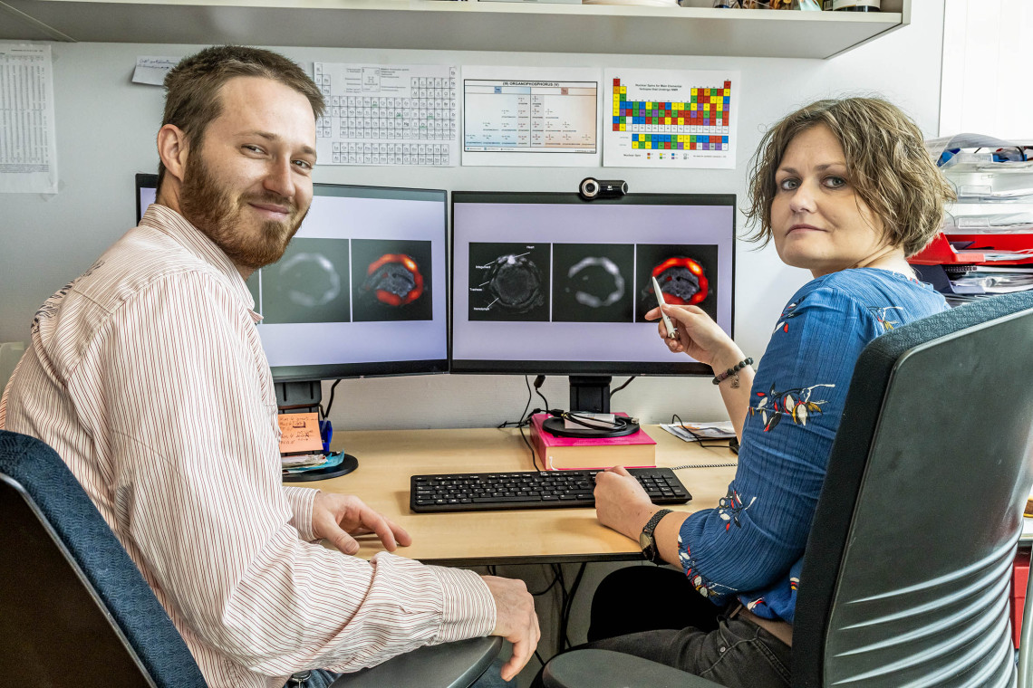

The images on the computer screen looks somewhat obscure to me: three black and white images of a circular shaped object, while the last image shows an obvious red color. ‘These are MRI images of a cross sections of a caterpillar’, Koshkina explains. ‘The red color represents our new molecule and shows how it has evenly spread in the caterpillar’s body.’
'We need to develop new agents that are both stable, but also biologically safe and environmentally friendly'
And although the caterpillar is the first patient illustrating the MRI application of the new molecule, over time, the scientists expect it will not only serve as a sustainable MRI contrast agent in humans, but also play an important role in targeted drug delivery and improved coatings for medicine.
Fifty shades of grey
MRI imaging technology has revolutionized the medical sciences by creating high-resolution and very detailed images of organs and tissues in the body. An MRI uses a strong magnetic field to create a picture showing different organs. Based on the amount of hydrogen (liquid) present in different body parts, the magnetic field creates images in over 50 shades of grey, visualizing the organs and tissues.
For example, fat tissue appears white, while tissues containing more liquid, like the liver, are darker. By injecting contrast agents in the patient, image detail can be increased even further. However, usually, contrast agents contain heavy metals and may accumulate in tissues and in the environment. ‘Such metal-based agents have been found in the brain of patients, but also in drinking water’, Koshkina says. ‘Therefore, we need to develop new agents that are both stable, but also biologically safe and environmentally friendly.’
Environmental advantages
Project partner Timo Rheinberger has been working on designing new molecules for quite some time in the group of Frederik Wurm before he joined Koshkina’s team. ‘It was fundamental, curiosity-based research, where I designed and built molecules based on phosphorus’, he says. Natural phosphorus (P) has in theory the right properties to be used as an MRI imaging agent, and could replace the current contrast compounds.
(Text continues below the picture.)
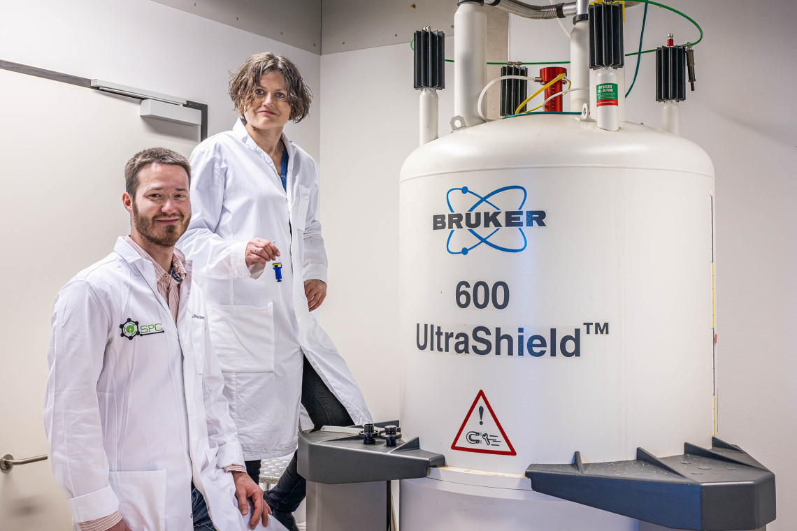
The expertise of Koshkina on polymer-based MRI agents and the extended knowledge on phosphorus-containing polymers from Wurm‘s team and Rheinberger’s thesis was a match made in heaven: together they successfully designed and built a completely new P-containing, non-toxic and biodegradable molecule, that could be used as an MRI imaging agent.
'Replacing the metal-based contrast agents, with our phosphorus-based polymers has huge environmental advantages'
Like any polymer, the long, chain-like molecule consisted of coupled, repeating units, monomers, with the P incorporated in the polymer backbone. ‘Replacing the metal-based contrast agents, with our phosphorus-based polymers has huge environmental advantages’, Koshkina says. ‘In addition, such a new molecule may also have several other applications in medicine.’
Overcome challenges
But the team had to overcome some challenges: background noise. P is abundantly present in the body and while MRI can detect this compound, it’s hard to distinguish the P in the contrast polymer from the natural body P, resulting in a lot of background noise and a less clear image.
To make the polymers more stand out against the P-rich background, the scientists had to pull a sophisticated trick from their sleeve. ‘Normally, a phosphorous group consists of P, surrounded by four oxygen atoms’, Rheinberger explains. ‘We changed the P group in the polymer slightly, by replacing one oxygen by a carbon atom. With this trick the MRI could easily distinguish between P in the body and polymer-associated P.’
Now, the background noise issue was solved, the scientists had to deal with the next challenge. The new P-containing polymer appeared less sensitive in MRI imaging compared to the traditional contrast agents. To solve this issue and increase the polymer visibility in the body, the scientists packed many polymer molecules together in a sphere-like shape: a so-called micelle: here the P density was very high and thus easily detectable by the MRI.
Breakthrough
But the scientists were not satisfied yet. To obtain the MRI image, and make the polymer stand out even more, scientist and project partner Uli Flögel, from the University of Düsseldorf, Institute for Molecular Cardiology, optimized the MRI method. They first made an anatomical black and white MRI image to show the organ positions. Then they made the same image using their P-based polymer as a contrast agent. ‘We sandwiched both pictures into one, using an algorithm, that showed the organs and made the locations of the polymer appear in red’, Koshkina explains.
‘It was a breakthrough that an ordinary MRI could clearly detect phosphorous in our polymer.’ Although the polymer has not yet been clinically tested, experiments on tobacco hornworm caterpillars proved very successful. ‘We injected caterpillars with polymer micelles and the molecules distributed evenly through the body and stayed there for more than 24 hours’, Rheinberger says. ‘This is a promising result and indicates the polymer is suitable for MRI imaging.’ Injection in the gut showed breakdown products in the caterpillar’s feces, indicating that the polymer was naturally degraded.
‘It was a breakthrough that an ordinary MRI could clearly detect phosphorus in our polymer’
Clear tumor image
Koshkina and Rheinberger have now set-up a spin-off company, Phos4nova B.V., to further develop their findings. An important aim is to bring the polymer to the clinic. A long process, and in the meantime, the company aims to improve the specificity of the polymer for certain organs or tissues, by attaching so-called targeting groups: a small molecule that binds specifically to certain tissues. ‘We are currently developing molecules that specifically bind to receptors that are only present in some types of cancer’, Koshkina explains.
‘Attaching such a molecule to our polymer, will result in binding of the polymer to the tumor receptor and consequently, a clear tumor image.’ Other applications of the polymer are encapsulating drugs and deliver them to, for example tumors, where they are released. ‘Tumors have higher metabolism and lack oxygen, while reactive oxygen radicals are present. Also, the pH inside tumors is lower’, Rheinberger says.
‘We can tune the encapsulating polymers in such a way, that they release the drugs when the pH is lower or when oxygen radicals are present, resulting in a very specific delivery of medication.’ While these applications are still future promises, they may drastically improve treatments of certain types of cancer.