Rouwkema has focused on tissue engineering his entire scientific career. His interest in vascular networks was sparked during an internship at MIT in 2002. ‘We were the first to demonstrate that it is possible to add vascular networks to other engineered tissues. Making that happen is one of the main challenges of getting engineered tissue to clinical practice,’ says the Associate Professor in the UT Department of Biomechanical Engineering, whose work on vascularization has earned him a research visit at Harvard Medical School, as well as the VENI fellowship and the ERC Consolidator Grant worth two million euros.
Macro and micro
Artificial blood vessels are of key importance if we ever hope to implant engineered organs in patients. ‘It might be the only way to get oxygen and nutrients to the tissue, which would otherwise die,’ explains Rouwkema. ‘To create a perfect vascular network, however, is extremely complex. It is a multiscale structure: there are bigger vessels branching into very fine veins and capillaries. The main challenge now is to bridge the gap between the macro and the micro structure.’
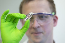
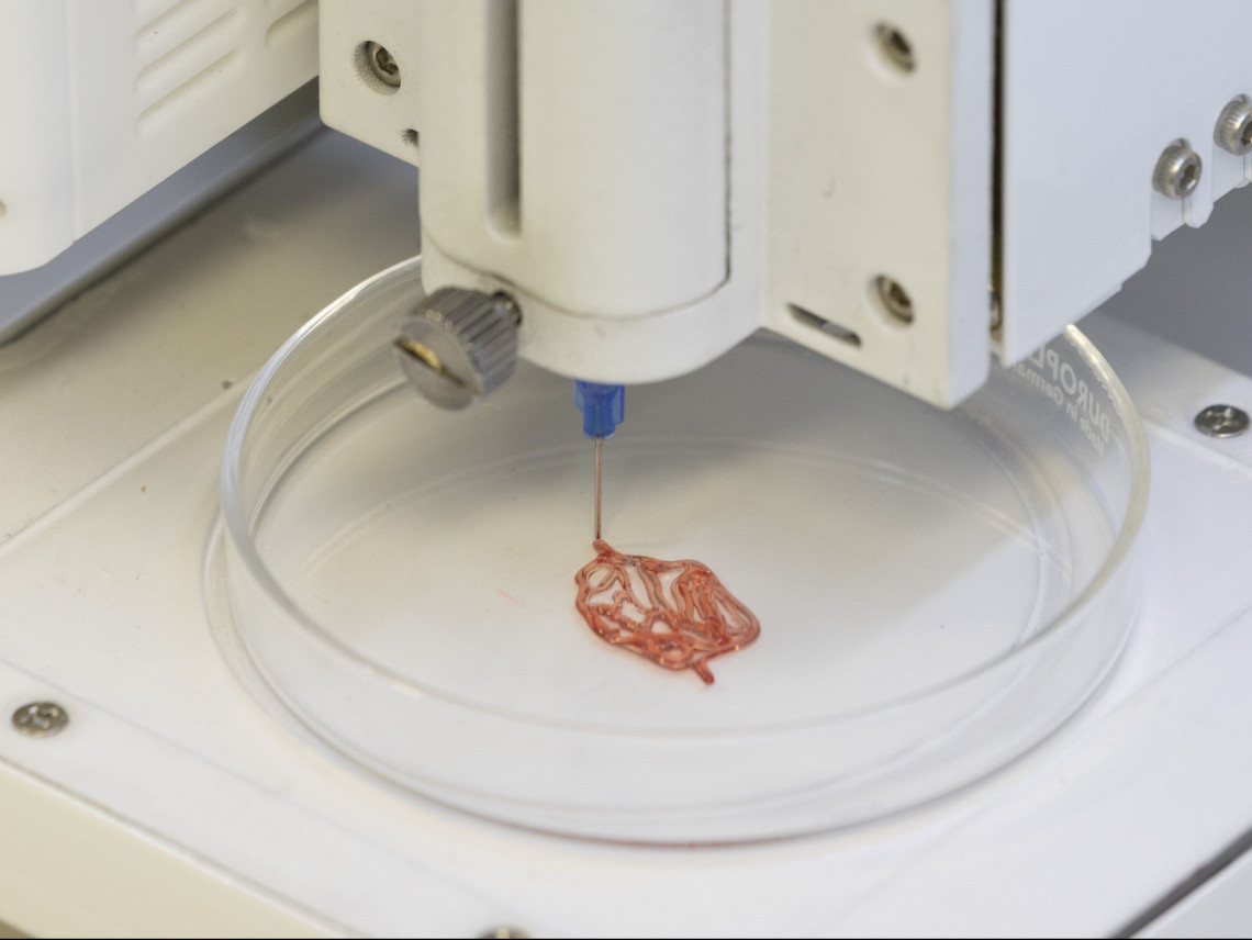
The larger structures can already be produced using 3D printing and similar biofabrication methods. What do these 3D printed tissues look like? ‘Jell-O pudding,’ answers the scientist. ‘We can essentially make small-scale Jell-O pudding in any shape with cells included inside. At first the structure is transparent but the cells start forming their own matrix, turning it opaque. We are able to print the shape of the tissue, such as cardiac tissue, and print a vascular network within it.’ Even ‘blood’ can run through these printed ‘veins’. ‘We use 3D printed structures composed of special sugar in the hydrogel. They dissolve in the watery environment, creating an empty cavity through which we can pump fluids.’
Steering nature
As Jeroen Rouwkema points out, the biofabrication of larger vessels is relatively well developed, but there is currently no fabrication method to produce the small capillaries. It might not be necessary to ‘fabricate’ these fine blood vessels, though. The UT researcher is testing a different approach.
‘We are using the natural organizational capacity of the vascular cells. Our biological system knows how to grow the vascular network. After all, it’s able to grow a whole human being from a single fertilized egg cell. We therefore try to use this natural capacity and steer the development using chemical and mechanical signals, moving it in the right direction.’ Understanding how to use signals to optimize the organization of vessels is the main goal of Rouwkema’s project VascArbor, for which he received the ERC Consolidator Grant in 2016.
To test exactly how vascular networks develop when various signals are applied, Rouwkema’s research group has started using chicken eggs. ‘We put the contents of a fertilized chicken egg into a transparent container, so we can follow how the vascular networks form around the yolk. The container also allows us to locally offer signals to the vasculature, to study how different signals regulate organization,’ explains the scientist.

Jeroen Rouwkema
2017-now Associate Professor in the Department of Biomechanical Engineering at the UT
2017 TERMIS EU Robert Brown Early Career Principal Investigator Award
2016 ERC Consolidator Grant
2014-2015 Visiting Researcher at Harvard Medical School
2014 Marie Curie International Outgoing Fellowship
2010 VENI Fellowship
2008 PhD in Vascularized tissue engineering, UT
2002 Internship at Massachusetts Institute of Technology (MIT)
Test animals and donor organs
‘Our current research is more fundamental. We first need to understand how we can control vascular organization before we can focus on applications,’ says Rouwkema. Yet, the real-life implications are on top of his mind. ‘There are two main applications of vascularized tissue engineering: drug testing and “replacement organs”. In the first case, the engineered tissue would be used in a lab for drug testing as an alternative to animal testing. Secondly, - in the long run – we could make the tissue from the patient’s cells, implant it into the body and ultimately replace donor organs.’
How far are we from making this a reality? ‘If it comes to drug screening platforms, for research applications we are already there. But to use it for the pharmaceutical industry would require standardisation and changes in legislation,’ answers Rouwkema. ‘Tissue implantation is already a reality, but only for simple and thin tissue, such as skin or cartilage. To be able to do the same with more complex tissue, we first need to solve several major problems, one of which is the implementation of vascular networks.’
'I hope that, at the very least, there will be no need for test animals twenty years from now'
Does the UT scientist believe this will be accomplished in his lifetime? Will we be able to create perfect vascular networks? ‘One of the big unknowns right now is determining how mature the tissue should be before we implant it. Until recently we assumed that it should be fully developed before we put it into the body. However, that might not be necessary. It might actually work better if we implant a tissue precursor that still needs to develop, so it can mature inside the body. If that is true, then engineered vascular networks might be less important because they would simply organize themselves in the body. It is hard to predict where the field will be by the end of my career, but I do believe my work is relevant. I hope that, at the very least, there will be no need for test animals twenty years from now.’