In the Netherlands, every day about 200 people are affected by cardiac arrest or a stroke, resulting in an acute blood and oxygen shortage in the brain. This is a life-threatening condition requiring immediate medical care to prevent death and minimize damage to the brain. ‘It is crucial to restore the blood supply to the brain as soon as possible,’ says professor Hofmeijer from the department of Clinical Neurophysiology. ‘Reanimation or removing a blood clot from the blood vessels are the first steps to minimize brain damage and increase the chances of survival and recovery.’
Unfortunately, even if a cardiac arrest patient survives after successful recovery, permanent brain damage may occur if the oxygen deprivation has lasted too long. After these first life-saving actions, hospital treatment to support and stimulate brain recovery are essential for an optimal recuperation of the patient. However, the reality is that such treatments are currently non-existent. ‘Comatose patients, after cardiac arrest, that have possible brain damage, are sedated and cooled down to calm the brain,’ Hofmeijer explains. ‘We tend to believe this promotes the brain healing, although this has never been proven. In fact, recent clinical studies suggest that this treatment has little to no effect.’
New therapies
Professor Hofmeijer is fascinated by what exactly happens in the brain after a period of oxygen shortage and how to apply the best possible treatment for an optimal recovery of the patient. Since it is difficult to study these processes and test new therapies for optimal recovery directly in people, she and her team use in vitro brain cell cultures to study the effects of oxygen shortage.
‘Brain oxygen shortage can be very acute’
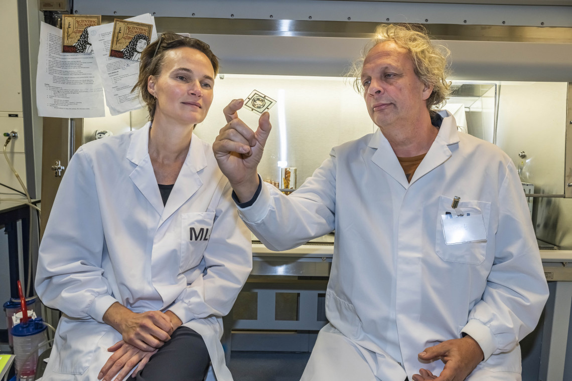
Hofmeijer’s colleague Joost le Feber is specialized in these lab experiments and together they investigate how brain cells respond to a lack of oxygen. ‘Brain oxygen shortage can be very acute, and in case of a stroke, part of the brain cells die immediately, for example in the center of an infarction,’ Le Feber says. ‘But around the dead brain cells, there is a zone where the brain cells receive just enough oxygen and nutrients to survive for a while. They may survive for as long as a day and with quick action, these could be saved, minimizing brain damage.’
Measuring brain activity in patients using an EEG shows that these brain cells, although not dead, show abnormal activity. Le Feber thinks this is due to energy shortage: the cell’s battery is empty. But restoring the oxygen and nutrient flow to adequate levels may result in brain cell recovery, where the communication between cells, through synapses, structures that pass electrical or chemical signals from one brain cell to the other, is restored.
Mimicking a stroke
To study these processes in more detail, the scientists use brain cell cultures, which can be obtained from rats or derived from human cells. For human brain cell cultures, first, human blood- or skin cells are reprogrammed to stem cells. With the right growth factors, these cells can be stimulated to differentiate into brain cells.
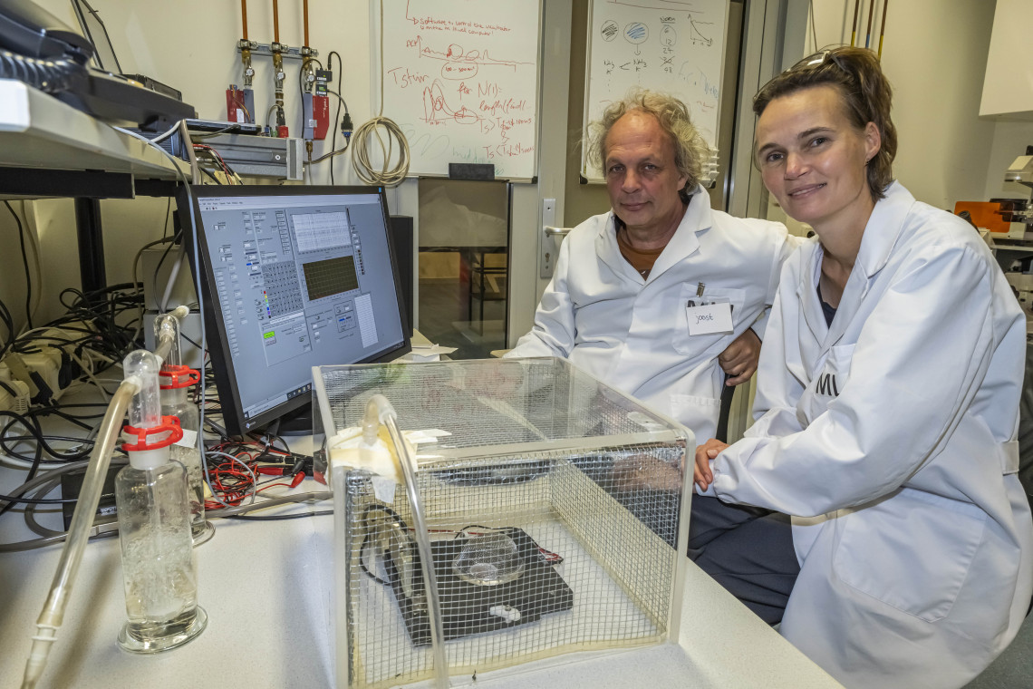
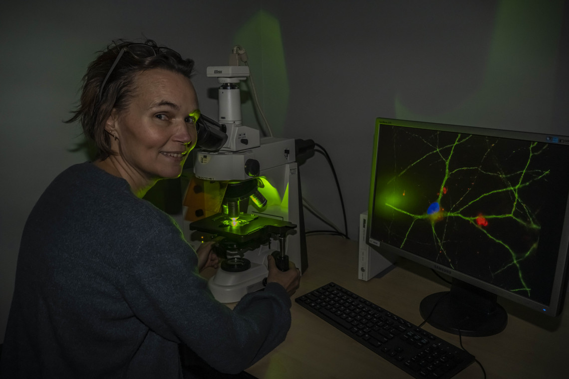
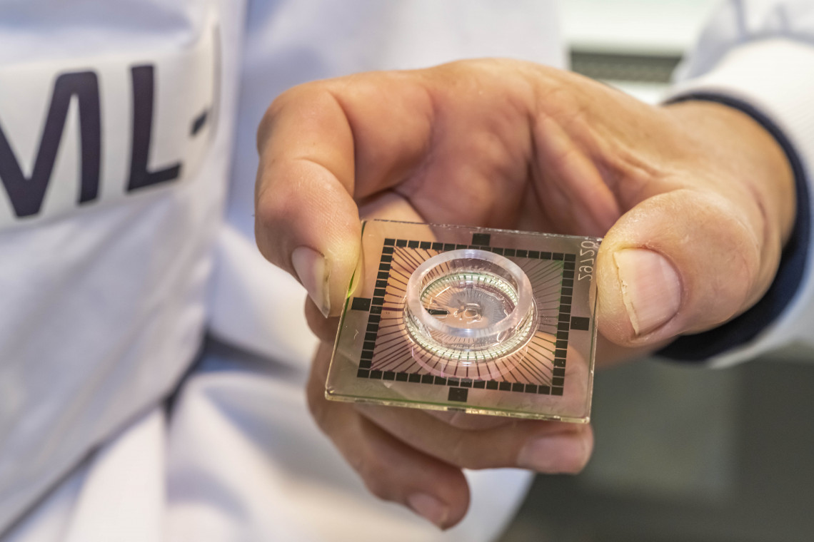 Le Feber points to his experimental set-up in the lab of Clinical Neurophysiology: ‘This is where the magic happens.’ Silicon hoses supply fresh air into a closed, Plexiglas box, where a small, round container holds living brain cells in a reddish fluid. ‘In this glass container, the brains cells attach to the bottom and start growing and connecting and communicating with each other, a process that takes about three weeks,’ Le Feber explains. ‘Electrodes on the bottom detect their activity, but they can also be used to stimulate the brain cells.’ When the brain cells have developed sufficient connections, they communicate, and activate each other, resulting in a synchronous activity. This activity is monitored by the connected computer, where a screen shows colored lights flashing synchronously. Le Feber: ‘This synchrony indicates that the brain cells communicate well and are healthy.’
During the experiments, the brain cells are subjected to different levels and durations of oxygen shortage, mimicking a stroke. A transient 100 percent oxygen deprivation simulates cardiac arrest. During the oxygen deprivation, the brain cell’s activities are continuously minored. Le Feber: ‘We have found that within hours after we decreased the oxygen supply, the brain cells start to decrease their activity and lose most of their synchronicity. With a minimal amount of remaining oxygen, such as in the border zones of a stroke, only after half a day to a day, cells eventually die, often by the so-called programmed cell death, a mechanism where cells die in controlled way, so they don’t damage the surrounding cells.’
Improved survival
The scientists hypothesize that programmed cell death is directly caused by the lack of brain cell communication. A bold idea, that challenged the current consensus that calming down and sedating the brain would improve recovery and minimized brain damage. However, their laboratory experiments confirmed their hypothesis: mild stimulation either electrically or by stimulating chemicals, like the neurotransmitter acetylcholine or appetite hormone ghrelin, improved the survival of oxygen deprived brains cells.
‘Our results suggest that in some cases a different treatment, like mild brain stimulation, is more effective’
‘Our results suggest that in some cases a different treatment, like mild brain stimulation, is more effective,’ says Hofmeijer. The lab results have been translated to a study in patients in the clinic, where the first trial on 160 patients with brain damage after cardiac arrest is currently carried out. Half of the patients received a mild brain stimulation by ghrelin, while the other half was treated in the conventional way by calming down the brain. The recovery of all patients is closely monitored during half a year.
Different points of view
The success of Hofmeijer’s and Le Feber’s research can likely be attributed to a combination of clinical and laboratory research. Hofmeijer is indeed convinced that to improve treatments, these different disciplines should collaborate. Therefore, she created a new Chair ‘Translational Neurofysiology’, where she puts this vision into practice. According to the professor, this requires a close collaboration between lab researchers and medics, where both parties learn to understand and trust each other. An important advantage is that the scientists involved develop a broader perspective and reinforce each other, while increasing their understanding. But Hofmeijer is also aware of possible bottlenecks that need to be addressed.
‘Combining these different vantage points is key to improve treatments’
‘Medics aim to find knowledge for a better treatment of patients, and often are less interested how it exactly works,’ she says. ‘Lab researchers on the other hand are very much focused on mechanisms and how and why things exactly work, and are less interested in the potential impact on patients. Combining these different vantage points is key to improve treatments and discover new points of view.’ This multidisciplinary approach creates a win-win situation where both science and patients benefit and may in this specific case be a first step to a better treatment of patients suffering from brain damage due to oxygen shortage.