‘This is how a typical joint fluid sample from a patient with gout looks under the polarization light microscope.’ Tim Jansen, rheumatologist at the VieCuri medical Centre in Venlo, points to a computer screen where beautifully arranged blue and yellow swirls are visible. It looks like an abstract painting of a gifted artist. ‘These are probably sodium urate crystals that are associated with gout,’ he explains. ‘But although this is a rather clear example of gout, in many patients, we can’t really be sure if the crystals we see consist of sodium urate. It might also be another type of crystal, associated with a different disease.’

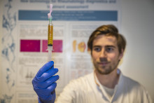
A few minutes later, we move to another room, where PhD candidate Tom Niessink (UT Medical Cell Biophysics group) shows a brand-new piece of equipment: the Raman spectroscope integrated with a polarization microscope. This machine might be a huge step forward in diagnosis of gout and other crystal-related diseases. Jansen and Niessink collaborate in their research to evaluate the added diagnostic value of the Raman method. Niessink takes the sample he and Jansen just analyzed under the polarizing microscope and places it in the machine. Within a minute, a colorful, wavy graph with some distinct peaks appears on the computer screen. ‘This is a spectrogram of the sample, and it has the characteristics and pattern of a sodium urate crystal,’ Niessink says. ‘Look at these high peaks, those are very typical for urate crystals. This patient definitely has gout. Diagnosis confirmed!’
Extremely painful
Gout is an auto-inflammatory disease, caused by a metabolic disorder, giving rise to high concentrations of uric acid in blood. Eventually, uric acid crystallizes and sodium urate crystals can be deposited in the joints. These crystals may cause heavy and extremely painful inflammatory reactions. Gout affects about two to five percent of the population, and is increasing.
‘Gout is usually diagnosed with a polarization microscope, that identifies the presence of crystals in a sample of joint fluid,’ Niessink says. ‘However, the crystal’s chemical composition usually is more difficult to determine using this method, and the sodium urate crystals may easily be confused with, for example, glass slivers or pyrophosphate crystals, the latter associated with pseudo gout.’ This disease has similar symptoms as gout, but it has a different cause and requires a different treatment. Using just the polarizing method to correctly diagnose gout is sometimes challenging.
Wrong diagnosis
Matthijs Janssen, former rheumatologist at Rijnstate Hospital in Arnhem, is expert on the limitations of the polarization microscope to diagnose gout: Since the 1980’s he has been using the polarization method to identify sodium urate crystals, and this requires a lot of training and experience. ‘The subjectivity of the human factor is a problem and although we can see the presence of uric acid and pyrophosphate crystals, there often is doubt which crystal we are dealing with,’ he says. ‘This may sometimes lead to a wrong diagnosis and treatment.’
‘This may sometimes lead to a wrong diagnosis and treatment’
To improve the crystal-based diagnostics for gout in patients, Janssen participated in a national competition among students from different universities about ten years ago. Several UT students came up with a promising solution: using a so-called Raman spectroscope in combination with the polarization microscope. First, polarization identified the presence of crystals in the sample, whereafter the Raman spectroscope analyzed their exact chemical composition by making a so-called spectrogram.

‘Raman emits light on the crystal, that is subsequently scattered in a pattern, unique for the crystal’s composition,’ Janssen explains. ‘The scatter pattern is subsequently transformed into a spectrogram, a graph containing different peaks, that corresponds with the carbon, oxygen and nitrogen atoms present in the crystal.’ Raman thus gives an exact fingerprint of the molecules present, and identifies the crystal’s chemical make-up. In addition, scientists can now also identify more different crystals, like calcium-containing crystals. This is not possible using just polarization. The new device, essential in this research project, is developed by UT Spin-off company Hybriscan Technologies B.V., under management of UT professor Cees Otto of the Medical Cell Biophysics group.
Added value
Niessink’s research, supported by ReumaNederland, is mainly focused on clinical validation of the method. This is crucial before the method can be used routinely. ‘I am currently examining samples of patients that are diagnosed with gout with 100 percent certainty. We use these samples to calibrate and evaluate the Raman spectroscope in combination with polarization,’ Niessink says. ‘When there is uncertainty about the diagnosis, Raman can be of important added value.’
‘When there is uncertainty about the diagnosis, Raman can be of important added value’
During his validation studies, Niessink also found crystals of unknown origin in joint fluids. For example, some samples contained previously unknown crystals, and also inorganic particles such as titanium oxide were found. This compound is used, among others, as a white coloring in cosmetic products. ‘These crystals might come from tooth paste,’ Niessink says. ‘Clearly, the method offers a wealth of possibilities to detect also crystals or particles that are associated with different diseases or even with pollution.’
Diagnosed more effectively
Therefore, besides validating the new method, Niessink is now also extending the application of the Raman method to examine a range of other diseases than gout. ‘Together with Maastricht UMC, we are currently examining joint fluid from a large sample bank to determine a possible role of crystal formation in patients with arthrosis. We suspect that this may play a role in the progression of this disease,’ Niessink explains.

But the presence of crystals, fibers or particles in tissues may also be associated with a specific job or even pollution. Examples of these profession-related diseases are asbestosis, where asbestos fibers accumulate in lung tissue, and silicosis, caused by inhaling large amounts of crystalline silica dust, present in rocks and concrete. And to the scientist’s surprise, they discovered microplastics in some samples. Also in these cases, Raman could help to diagnose the problem more effectively. Another possible application is in breast cancer research. Niessink: ‘In progressing breast tumors the inner part of the tumor dies off due to insufficient nutrient supply, and microcalcifications are formed. This process can also be examined with the Raman technology. In fact, any tissue where some kinds of crystals are formed, can be studied with this method, and we can even apply it to environmental research to microplastics. The potential is enormous!’