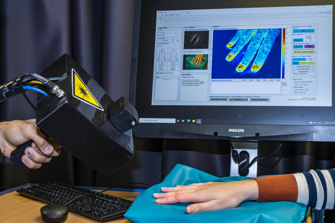
‘Look how our newly designed handheld LSCI makes this beautiful blood perfusion image,’ scientist Ata Chizari says. He points the 20 by 20-centimeter black laser device, nicknamed HAPI, handheld perfusion imager, to the hand of master student Technical Medicine Sacha Teunissen. A laser beam illuminates her hand and on the computer screen, a hand-shaped image appears in blue, yellow and reddish colors. The redder, the higher the blood flow. Chizari: ‘This new design is much smaller and easier to use compared to the existing LSCI technology. It can therefore be used for more clinical applications.’ To compensate for the hand-induced movement, the researcher has adapted the laser beam and applied image stabilization using smart software.
Powerful tool
For several decades, the Laser Speckle Contrast Imaging (LSCI) technology has been a powerful tool in medical diagnostics by measuring relative blood flow in tissues. The applications of this technology are numerous, for example in assessing the severity and depth of burn wounds. Since badly damaged and dead tissue will not show any blood flow, measuring the perfusion in the burnt tissue using LSCI, gives accurate information regarding the gravity of a particular burn. Also in the skin disease psoriasis, the growth of lesions can be assessed by measuring relative blood flow using this method, since perfusion is higher in these lesions than in healthy tissue.
Blurring effect
The LSCI technology uses a laser beam to illuminate tissue, for example skin. The reflected light forms a random interference pattern, the so-called speckle pattern, consisting of dark and bright areas. ‘When skin is illuminated by a laser beam, the light interacts with moving red blood cells inside the capillaries of the skin tissue, and this causes the speckle pattern to continuously change in intensity,’ Chizari explains. ‘An image of such a dynamic speckle looks blurred. The higher the blood flow, the more blurred and less contrasty the speckle image appears.’ Physicians measure this blurring effect using photodetectors, like CCD or CMOS sensor cameras, and computer software turns this into a colorful blue, yellow and red image, showing the relative tissue perfusion.
Lack of flexibility
However, the current commercially available technology is cumbersome and lacks a convenient operation. For example, due to its bulkiness and inflexibility, it requires difficult maneuvers and body positions when measuring on areas such as knees and elbows. Also, due to the size alone, its use in operating rooms is limited as well. Especially during transplant surgery, like breast reconstruction after surgical removement of a tumor, LSCI may supply the surgeon with crucial information of the blood supply of the newly transplanted tissue during and after the actual operation. Chizari: ‘The equipment may also be intimidating for young children. By using a portable, hand-held device, the flexibility and practical use of the technology would increase substantially, addressing these problems and extend its use to a broader clinical situation.’
‘By using a portable, hand-held device, the flexibility and practical use of the technology would increase substantially’
Unreliable diagnosis
The past four years, the team has been working to design a hand-held version of the traditional LSCI device. ‘The main challenge was to compensate for the hand movements, while holding the device,’ Chizari says. ‘This movement has two problems: it causes extra blurring of the speckle images, resulting in an unreliable result that suggests more blood flow in the tissue. In addition, this movement makes it difficult to follow a region of interest when recording a number of frames.’
To minimize the effects of hand movement, it matters how the wave front of a laser beam is shaped, Chizari and his team found out, after he tested the sensitivity of various, differently shaped laser beam fronts to movement. These laser fronts can be modified by directing the beam through a series of lenses. ‘We compared so-called scrambled wave fronts, used in commercial LSCI devices, with planar and spherical shaped ones’, the scientist explains. ‘The commercial scrambled beams were the most sensitive to hand movement and consequently, resulted in the largest movement artefacts. Modifying the laser wave front to either a planar or spherical shape resulted in a much better result.’ The team filed a patent for this uncomplicated hardware modification.
Smart data processing
Now the blurred speckle image problem due to movement in the hand-held device was diminished, Chizari and his team needed to deal with the second problem that occurs due to movement in the hand-held device: the difficulty to properly follow a specific region of interest in a number of subsequently captured frames. To solve this, Chizari made use of black marked points on the tissue, made by clinicians, that served as position indicators. Then, he deployed a post-processing algorithm that was able to detect the hand-held movement by tracking these markers and subsequently compensate for the movement.
‘Our hand-held device performs almost as good as the commercial devices,’ Chizari says. ‘On average, we measure a 16% higher blood perfusion in the hand-held device due to movement. This might be small enough to use this smaller device as a reliable diagnostic tool.’
‘Our hand-held device performs almost as good as the commercial devices’
Now that the main hurdles for a reliable hand-held LSCI have been taken, the scientist continues to further develop the performance of HAPI by applying a better image processing algorithm that can track the movement without having black markers on the tissue. In addition, Tom Knop, the research engineer of the team, is working on developing a wireless version that is even more flexible and user-friendly. Chizari: ‘With a wireless machine, the surgeon has even more possibilities to visualize the blood perfusion during an operation. That is a huge advantage in transplant and reconstruction surgery for the detection of complications that are not visible to the naked eye.’