The method also offers a non-invasive tool to characterize the tumor, and consequently a customized cancer treatment. Last year, their company ECsens won the prestigious 4TU Impact Challenge, a student innovation competition between the technical universities.
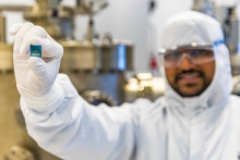
The clean-room of the University of Twente is a magical place. A sterile looking, dust-free, white environment, where people in protected suits, walk among impressive machines. This is where the magic happens: this ultra-clean space is the central laboratory for micro- and nanofabrication for scientists and the place where Mathew built his ground-breaking sensor technology.

In a yellow-lit room, Dilu Mathew, all covered up in a dust-free suit, slides a silicon wafer, containing multiple sensor chips, in a holder and places it inside a so-called electron-Beam Lithography machine. ‘This is the step that creates the smallest features of our sensor,’ he explains. ‘Inside this machine the necessary nanostructures are patterned onto the chip.’ After this last procedure, the chip is ready to detect any tumor fragment present in the blood of patients.
Tumor activity
Cancer still is one of the leading causes of death worldwide, second only to cardiovascular diseases. Every year, there are about 17 million new cases of cancer, while about 10 million cancer patients die each year, the majority due to metastatic cancer. Cancer may spread in the body when tumor cells become dislodged and enter the vascular system. The number of these circulating tumor cells are indicative for the tumor activity and long-term survival. However, measuring these circulating tumor cells to track down metastatic cancer is challenging, because cancer cell concentrations are very low. ‘In one milliliter of blood there can be about five billion red blood cells, and only one tumor cell,’ Pepijn Beekman says. ‘Measuring this single cell among this vast number of red blood cells is a problem.’
Accurate method
Luckily, the blood may contain more biomarkers for tumor activity than intact circulating tumor cells. Tiny cancer cell fragments also may end-up in the circulation and provide similar information regarding survival and tumor activity as the number of circulating tumor cells. ‘These so-called tumor-derived extracellular vesicles, tdEVs are more than 10.000 times more abundant than tumor cells, and therefore easier to find,’ Dilu Mathew explains. ‘Therefore, we focused our research efforts on developing a sensitive and accurate method to detect these fragments.’ Eventually, the scientists managed to develop a tiny sensor for accurate electrochemical detection of tdEVs present.

On a nanoscale, the basic design of the sensor looks like two electrodes, separated by a gap of only 100 nanometers. That is 400 times smaller than the thickness of a human hair. The whole sensor consists of many of those electrode sets, resembling two combs, with the teeth pushed together, leaving a 100 nanometer gap in between. On each of the electrodes, antibodies are attached, that are specially designed to capture tdEVs. This antibody is highly specific for tumor cell fragments, since it only binds to certain proteins, so-called antigens, that are only present in tdEVs and not in blood cells.
‘One grain of rice in an Olympic-size swimming pool’
‘When blood is flushed over the sensor, the tdEVs are recognized and captured by these antibodies,’ Beekman explains. ’In a next step, a fluid containing a second antibody, a so-called reporter antibody, is flushed over the sensor. This antibody also binds to the captured tdEVs, and translates this into a signal through a cascade of chemical reactions which can be measured electrochemically.’ The fact that the sensor is incredibly small helps to amplify the signal. ‘Right now, we are able to measure 10 tdEV particles in one microliter of sample,’ says Beekman. ‘That is comparable to one grain of rice in an Olympic-size swimming pool.’
Multiple applications
According to the scientists, their new sensor has multiple applications. First, it can be used in diagnostics: by measuring the tdEVs, the sensor can prove the presence of a tumor and in addition, say something about its activity. A more promising application is its use in finding the best treatment for a certain tumor. Different antibodies are developed that can only bind to one specific tumor type. By investigating to which tumor tdEVs a specific antibody binds, the scientists can detect and characterize the tumor present. If the tumor type is identified, the best treatment for that specific tumor can be selected. The effectivity of the chosen treatment can then be assessed by monitored the number of tdEVs, and thus tumor activity before and after treatment.
‘This is an important step in cancer therapy,’ Beekman says. ‘New cancer treatments, like immune therapy, can be very effective, but work only in 20 percent of the cases. By assessing the tumor type, we can better predict if immune therapy will work.’ Eventually this makes cancer treatment more cost-effective.

Clinical trials
The last year the scientist have improved their technology. The sensitivity has increased significantly: now the scientists are close to detecting a single tdEV in one milliliter of blood. This means that instead of measuring tdEV concentrations, they can now simply count the number of these tumor fragments, and get the results directly, in real time. ‘To make the technology ready for use, we have started to test the clinical proof of concept using patient samples together with Deventer hospital,’ Mathew says. ‘We expect the results by the end of this year. If this is successful, we will start clinical trials, using larger numbers of patient samples.’ A long and costly process, but worth the efforts, because the new sensor is more cost-effective and less invasive than imaging techniques, such as MRI: a simple blood sample is enough to get detailed answers regarding tumor type and activity.
Despite their success, the scientists remain modest and level-headed: ‘Most of the time, research is a lot of struggle, with many problems to be solved,’ says Beekman. ‘We feel very lucky and blessed that we have some moments of glory and it all has worked out.’