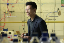
‘This is our newest batch of living heart muscle cells,’ Robert Passier says, while holding a 15-centimeter wide plastic rectangular dish, containing six different wells. Each well holds a reddish fluid with dissolved nutrients, nourishing the heart cells. The heart tissue is visible as an opaque film on the bottom of the well. Under the microscope, the film reveals a spectacular picture: it consists of thousands of heart tissue cells, that are closely connected to each other. They even behave like a real heart and about every few seconds they contract harmoniously. Just a few weeks ago, these pulsating heart muscle cells were human stem cells.
‘Since 2007 researchers are able to create so called pluripotent stem cells from human tissue, such as skin cells, blood cells or even cells from urine,’ Passier says. ‘These stem cells have the same properties as embryonic stem cells: in theory, they divide forever and can be grown into any tissue. In our case, we make them develop into heart muscle tissue.’ Converting stem cells into heart muscle cells requires quite some knowledge about genes: the pattern of active and silent genes determines what tissue the cells will form. Passier and his team know which genes need to be switched on or off to transform stem cells into heart muscle cells. The scientists regulate this specific gene activity pattern using, for example, special proteins that modulate the activity of individual genes.
Enormous breakthrough
The novel stem cell technologies are an enormous breakthrough and continue to develop. However, one of the challenges the scientists face is the immaturity of the heart cells formed from stem cells: they resemble foetal rather than adult heart cells. Understanding how to promote the ripening of these foetal cells into mature heart tissue is important because the in vitro cultures used for research should be comparable to adult heart cells. Therefore, one of Passier’s studies focuses on how different molecular markers, like DNA and RNA, change during the ripening from foetal to adult heart tissue cells. ‘We expect to see patterns during the ripening process,’ Passier says. ‘For example in gene activity: which genes will be switched on or off during the conversion from foetal to adult heart cells?’
Fix the genetic defect
Besides heart cell ripening, Passier and his colleagues focus their stem cell research on learning as much as possible from heart diseases and their treatment. With developing technologies, they can now make stem cells from patient tissue, for example from patients suffering from cardiomyopathy. This is a life-threatening heart disease with a strong genetic component. Stem cells developed from tissues of these patients contain the same DNA defect causing the disease. They often reflect the disease characteristics in a reliable way. This allows scientists to study such hereditary heart diseases in vitro. Passier: ‘But we can also imitate a disease in our cultured heart cells by mutating the stem cells, thereby mimicking the genetic defect, before we let them grow out into heart tissue.’
'We can imitate a disease in our cultured heart cells'
Testing heart medication also is an important research topic. For instance, checking the toxicity of newly developed drugs, or testing the effects of medication on heart cell functionality. ‘It is possible to measure the electrical activity of cultured heart tissue with heart rhythm disorders (comparable to an ECG in patients) and subsequently study the effectivity of new medication,’ Passier explains. ‘But we can also try to fix the genetic defect in cell cultures with a hereditary disease, using a state of the art technology like CRISPR-Cas. Afterwards, we can check the success of the procedure.’ All these investigations can be performed using the patient’s own tissue cells, that are converted into stem cells and subsequently transformed into heart tissue cells. Therefore, the experiments are highly reliable and mimic the real-life situation.

Perfect in vitro model
The efforts of Passier and his team to make in vitro cell cultures more and more realistic is gaining momentum due to the diverse expertise present in his team. Scientists from Twente, Nijmegen (Radboud University), Leiden (LUMC) and Amsterdam (AMC, VUMC) join forces to develop the close to perfect in vitro model by combining different disciplines in the fields of stem cell and molecular biology, heart development and disease, and tissue engineering. An important step towards almost life-like in vitro tissues is the development of so called organ on chip. Heart muscle cells can be grown in minute channels, milled out in a polymer material, for example. An artificial, miniature stream of medium supplies nutrients and oxygen to the cells. ‘Using this technique we can study the cell processes live under a microscope,’ Passier explains. ‘We can see how the cell divides, if it survives or dies and how it functions.’ To get a step closer to a real organ, different cell types can also be grown in the different channels, enabling scientists to study the interaction between different cell types, just like in an organ.
Real life situation
The development towards 3D tissue models is another important step towards real cultured organs. This is a huge and complex technological achievement, but it allows to study different cell types and their interaction, just as in a living human. ’Heart tissue has many different cell and tissue types, like atrium and ventricle muscle, pacemaker cells, blood vessel cells, and connective tissue,’ Passier explains. ‘We can make all these individual cells from stem cells and put them together. When conditions are right, they organize and interact with each other in a realistic life-like way.’ An important advantage of this technology is that the use of experimental animals can be greatly reduced. However, scientists have to prove that the new in vitro models perform as well, or better, than in living animals.
'We might be able to regenerate tissues and organs in the lab for transplantation'
‘Using these 3D models, we hope to effectively and reliably study diseases, but also applications towards the regenerative medicine are possible,’ Passier says. ‘If we know how cells grow and interact with each other, we might be able to regenerate tissues and organs in the lab for transplantation in the patient in the future.’ These scenarios are still far away, but the research is progressing fast. ‘I’m really happy with this team. Due to our different expertise, we complement each other. The project is expanding like a snowball.’
You can also find the interview in our latest Science & Technology Magazine. Grab a printed copy at the campus or see the full magazine online.


